
We have developed novel PINK1 biosensors to explore the biology of this important mitophagy regulator and for therapeutic development.
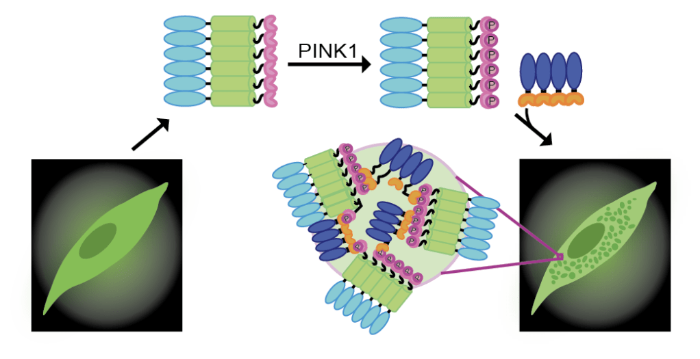
PINK1 is a mitochondrial serine/threonine kinase involved in mitophagy, the selective degradation of damaged mitochondria. Active PINK1 phosphorylates ubiquitin and the E3 ubiquitin ligase Parkin, resulting in the ubiquitination of outer mitochondrial membrane proteins and subsequent degradation of the damaged mitochondrion by autophagic machinery. Loss of function, autosomal recessive mutations in PINK1 are known to cause early onset Parkinson’s disease (EOPD). To better understand the role of PINK1 in mitochondrial function and towards development of therapeutics for EOPD, we have developed a novel biosensor for PINK1, called PINK1-SPARK.
Functionality of the construct relies on three components. The first is kinase sensing, allowed by the recognition of a short substrate peptide for PINK1 derived from ubiquitin by a phosphoamino acid binding domain (FHA2). Secondly, multivalency allows for liquid-liquid phase separation of the construct, conferred by homooligomeric tags (HOTags) that associate to form hexamers and tetramers in vivo. Finally, fluorescence signal (provided by enhanced green fluorescent protein, EGFP) allows for visualization of PINK1 activity. Upon PINK1 activation, PINK1 phosphorylates its ubiquitin-derived substrate peptide which is then recognized by FHA2. The resulting proximity of multivalent homooligomeric tetramers and hexamers promotes phase separation and visualization of PINK1 activity as green fluorescent puncta.
With PINK1-SPARK, we are able to measure endogenous PINK1 activity in single cells. PINK1-SPARK has been used in multiple cell types, with various PINK1 activating conditions. You can read about PINK1-SPARK here. We are now using PINK1-SPARK to study PINK1 in the context of EOPD and identify novel PINK1-activating small molecules.


Leave a Reply